Laboratory Biology

As a research biologist, I have used both empirical experiment and computer simulation in my Evolutionary Developmental Biology ("Evo-Devo") research, studying the development of embryos and body plans and the evolution of the processes that give rise to them. My main experimental tools have been embryo microsurgery and morphological microscopic analysis. Below I present images and video demonstrating both of these, followed by a microscopy lecture I gave, and my research publications. For more details, see my resume (download [industry style Resume] [academic style CV]).


Jump to:

Microscope images
Video clips: Embryo microsurgery
Video: Lecture on histograms and LUTs
Selected publications


Microscope images

(Click each image to view a
larger version in a new window.)

Of the many images I have produced over my years in embryology research, these are some of my favorites. They cover a variety of microscopy techniques and several organisms.


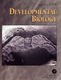
Journal cover for my 1996 paper.

Scanning Electron Microscopy (SEM).

Cross-section through a frog embryo just beginning neurulation (neural folds, notochord, somites, archenteron, with individual cells visible). Distance across: somewhat under half a millimeter.


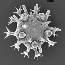
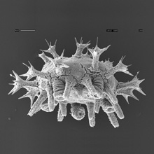
Scanning Electron Microscopy.

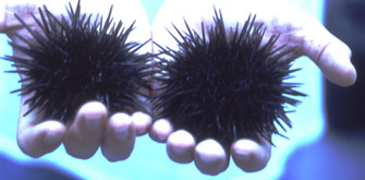
Tiny juvenile (newly metamorphosed) sea urchins – a little over half a millimeter across, showing their 5-part radial symmetry. (Note the 5 tiny round "tube feet" arranged in a pentagonal pattern in the first image.) Also note the two distinct kinds of spines: the blunt ended or "adult" spines, and the splayed or "juvenile" spines. For comparison, some adult urchins are shown below.

adult sea urchins
 

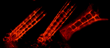
Polarized Light Microscopy.

The intricate skeleton in the spines of a juvenile sea urchin, viewed under polarized light, shines brightly through the transparent surrounding tissue because it is composed of inorganic crystals that effectively realign the vibration of the light to pass through the polarizing filters. Two "adult" spines left and center, "juvenile" spine right. (Compare to SEM images above.) See my 2002 and 2009 papers.


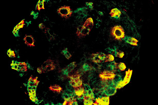
Confocal Microscopy with fluorescent antibody staining.

Juvenile (newly metamorphosed) sea urchin, stained with two fluorescently-tagged antibodies – one (red fluorescence) labeling a protein on the cells that secrete the skeleton, and the other (green fluorescence) labeling a protein within the crystal skeleton itself. Yellow indicates both proteins in close proximity. See my 2002 paper.


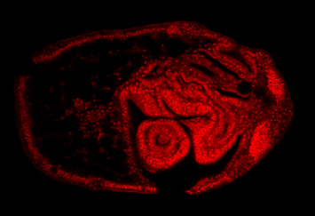
Confocal Microscopy.

Larval sea urchin, with the juvenile form (see above images) developing within it, almost ready to metamorphose. The ringed structure near the bottom is a cross-section through one of the tube feet. The stain is a fluorescent dye that labels the cell nuclei. See my 2002 paper.


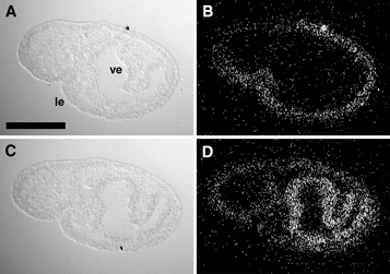
Dark Field Microscopy, for radioactive in situ hybridization.

Each photo is a slice of a larva like the one above. The two slices on the left are shown using DIC optics, and the ones on the right are the same two slices, viewed in dark field to reveal tiny silver grains. The grains were produced in a photographic emulsion by exposure to radioactivity. Radioactivity in a particular tissue shows that genes of interest were being expressed in that tissue. The slice in the top row, and the slice in the bottom row, are two nearby slices from within the same larva, treated to show two different genes. See my 2002 paper.

Reflected light, stereomicroscope image.

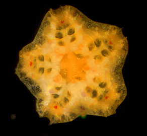
Juvenile starfish, a bit over a millimeter wide, 71 days after in vitro fertilization. The red spots are eyespots – light-sensitive, but not real eyes and they can't see actual images. Note the 5 arms, representing the same kind of symmetry displayed by its cousins, the sea urchins. Many tube feet are present, but at this stage are transparent and therefore difficult to see. (They can be seen more easily in the younger specimen shown below.)


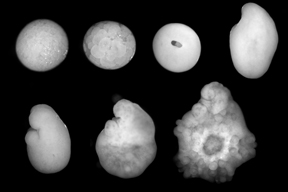
Composite image. Reflected light, stereomicroscope.

Seven successive stages of earlier starfish development, precursors of the juvenile shown above. Increasing in age from left to right and top to bottom. The first three specimens are embryos, and the last three are larvae, with the fourth being transitional between the two. Many tube feet are visible in the oldest specimen (lower right).


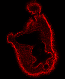
Confocal Microscopy.

Starfish larva at around the same stage as the 5th specimen (lower left) in the image above. Although the juvenile features (tube feet, 5 arms) are not yet externally visible at this stage, the confocal image allows us to see their earliest beginnings inside the larva. Note the internal cavity. At one end of that cavity, toward the bottom of the figure, you can see three projections. These represent 3 of what will be the 5 arms of the juvenile, after metamorphosis. The stain is a fluorescent dye that labels the cell nuclei.


Video clips: Embryo microsurgery

In 1996, I made a video for Xenotek Engineering, to advertise their "Gastromaster," a device designed to facilitate embryo microsurgery by using electrically generated water microcurrents to cut through tissue. These video clips show my embryo microsurgery in action – not to mention some really cool embryos, and of course a great product. (Click the images to view the movies in a new window. Video digitization provided by Xenotek Engineering.)


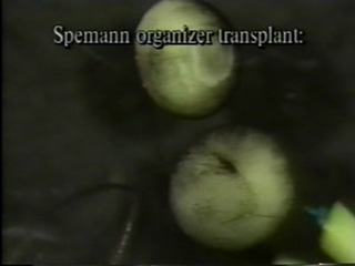
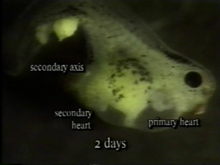
In this clip (50 sec, 8 MB) I perform the classic Spemann-Mangold organizer graft, inducing a second embryonic axis in an embryo of the frog Xenopus laevis. This involves transplanting the "organizer," a special tissue capable of inducing organ development in nearby cells, from one embryo to another. The embryos are about a millimeter wide, and the tools are hand-held. The tool on the left is made of a loop of my own hair. On the right would normally be my "scalpel," made of the same material; but here you see the Gastromaster instead.

After cutting both the recipient embryo and the donor embryo, I transfer the organizer tissue from the donor, into the pocket carved out of the recipient — but the actual transfer was edited out of the clip, so all you see is that suddenly I am tapping the transplant into place. After two days of development, an abnormal tadpole develops, with two hearts. Most obvious is the beating of the primary heart, near the head. To see the beating secondary heart toward the back, smaller and partially hidden behind other tissues, you might need to move the slider back and replay the very end of the clip a few times.

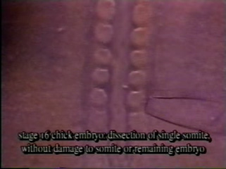
In this clip (15 sec, 2.5 MB) I use the Gastromaster to dissect out a single somite from an early chick embryo. This tiny structure is about a tenth of a millimeter wide.


Video: Lecture on histograms and LUTs


In fall 2008, it was my privilege to participate in teaching the first cohort of students in the brand new, one-of-a-kind Merritt Microscopy Program (MMP) at Merritt College in Oakland, CA. I taught Biosci 4: Advanced Microscopy Practicum (individual student research projects); provided individualized hands-on open-lab instruction; and, I gave the following guest lecture on histograms and LUTs in Biosci 3: Advanced Fluorescence/Confocal Microscopy.


Get Flash to see this player.


Part 1: Review of basic math fundamentals – graphing
and (generic) histograms. (22 minutes)

Get Flash to see this player.


Part 2: Images and image histograms. (27 minutes)

Get Flash to see this player.


Part 3: Introduction to histogram adjustment with LUTs
and gamma function. (24 minutes)

Get Flash to see this player.


Part 4: Gamma function, dynamic range, image optimization
pre- and post-capture, gamma. (44 minutes)

(Videos courtesy of MMP student Roger Castellanos.)


Selected publications


Minsuk, S. B., F. R. Turner, M. E. Andrews and R. A. Raff (2009). Axial patterning of the pentaradial adult echinoderm body plan. Development Genes and Evolution 219:89-101. [journal link] [download pdf] *

Minsuk, S. B. and R. A. Raff (2005). Co-option of an oral-aboral patterning mechanism to control left-right differentiation: the direct-developing sea urchin Heliocidaris erythrogramma is sinistralized, not ventralized, by NiCl2. Evolution & Development 7:289-300. [journal link] [download pdf] *

Minsuk, S. B., M. E. Andrews, and R. A. Raff (2005). From larval bodies to adult body plans: patterning the development of the presumptive adult ectoderm in the sea urchin larva. Development Genes and Evolution 215:383-392. [journal link] [download pdf] *

Minsuk, S. B. and R. A. Raff (2002). Pattern formation in a pentameral animal: induction of early adult rudiment development in sea urchins. Developmental Biology 247:335-350. [journal link] [download pdf]

Minsuk, S. B. and R. E. Keller (1997). Surface mesoderm in Xenopus: a revision of the stage 10 fate map. Development Genes and Evolution 207:389-401. [journal link] [download pdf] *

Minsuk, S. B. and R. E. Keller (1996). Dorsal mesoderm has a dual origin and forms by a novel mechanism in Hymenochirus, a relative of Xenopus. Developmental Biology 174:92-103. [journal link] [download pdf]


* I originally provided only journal links for these, because I do not legally have the rights to my own papers, which are available from the journals for a fee. After the death of Aaron Swartz, I decided to make these papers available here. See:
New York Times
slate.com
#pdftribute
pdftribute.net